Planmeca ProMax® 2D S3
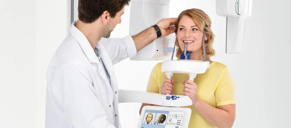
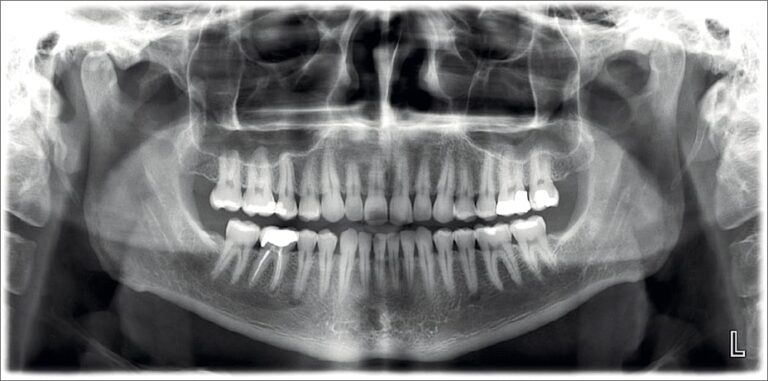
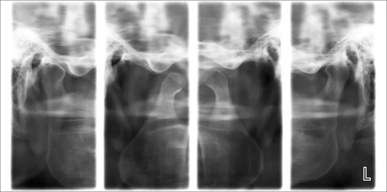
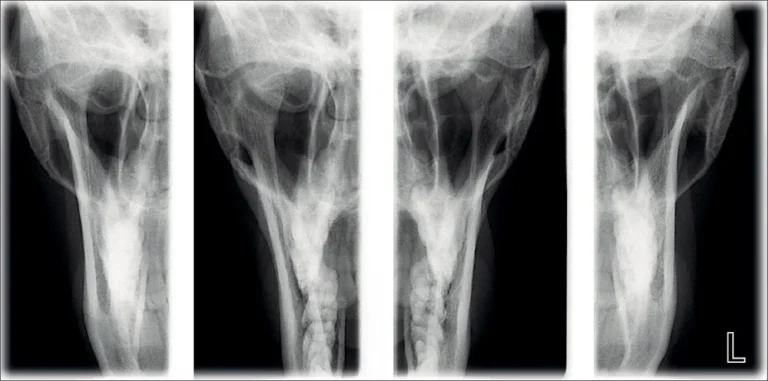
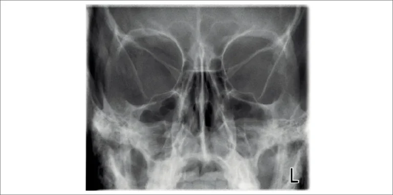
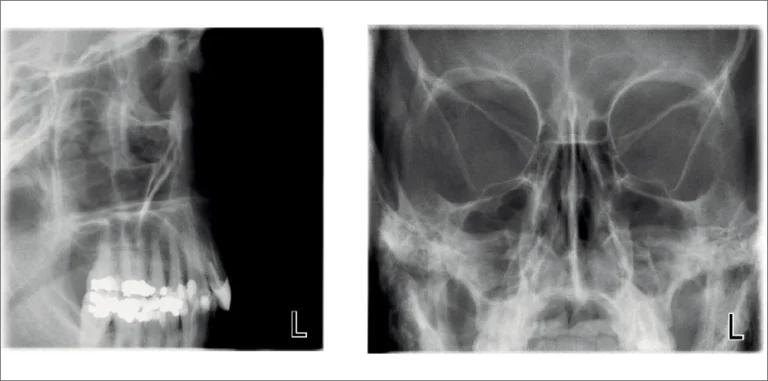
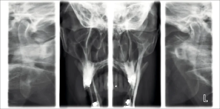
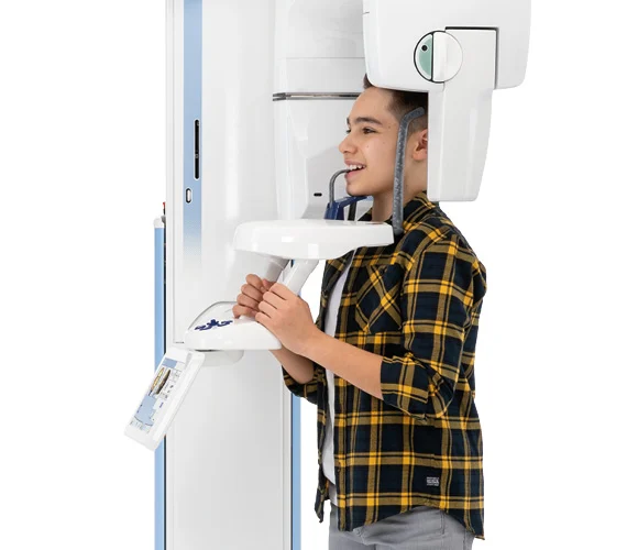
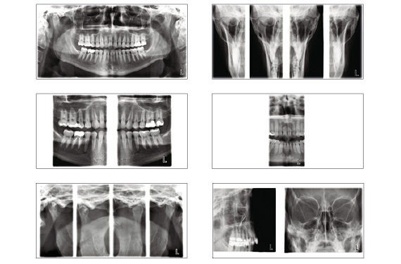
The three-joint (SCARA3) Planmeca ProMax 2D S3 unit has been designed for all 2D imaging needs: panoramic, true extraoral bitewing, TMJ and sinus imaging.
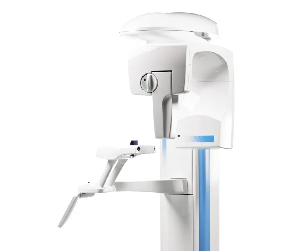
Planmeca ProMax® 2D S2
The two-joint (SCARA2) Planmeca ProMax 2D S2 panoramic dental imaging unit includes basic programs for panoramic, extraoral bitewing, TMJ and sinus imaging.

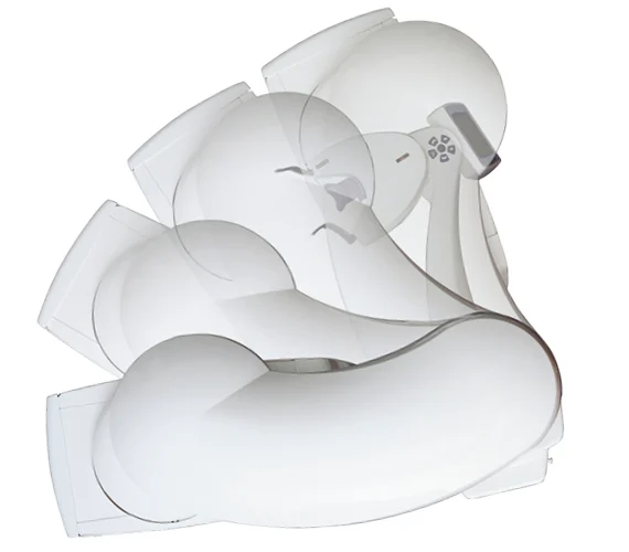
Robotic arm technology
Due to patented SCARA technology, our Planmeca ProMax units can produce all the required movements for rotational maxillofacial imaging. The Selectively Compliant Articulated Robot Arm guarantees an anatomically accurate imaging geometry – resulting in clear and error-free images.
Extensive imaging programs
The two-joint (SCARA2) Planmeca ProMax 2D S2 panoramic dental imaging unit includes basic programs for panoramic, extraoral bitewing, TMJ and sinus imaging.
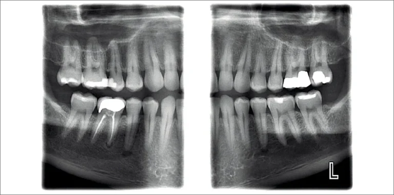
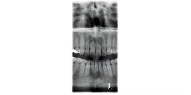
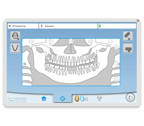
Perfect panoramic images with Autofocus
Our Autofocus feature for Planmeca ProMax® 2D S3 helps to guarantee consistently high panoramic image quality. It uses a low dose scout image of the patient’s central incisors to automatically position the focal layer. This dramatically reduces the need for retakes and guards the patient from unnecessary exposures.
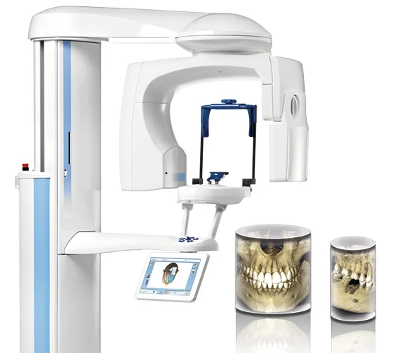
Fully upgradable from 2D to 3D
Planmeca ProMax 2D panoramic dental imaging units have been designed with upgradeability in mind. Their modular structure allows easy conversion to different imaging modalities. Whether you are upgrading your 2D unit to 3D or adding a cephalometric arm, Planmeca has the right solution for you.
