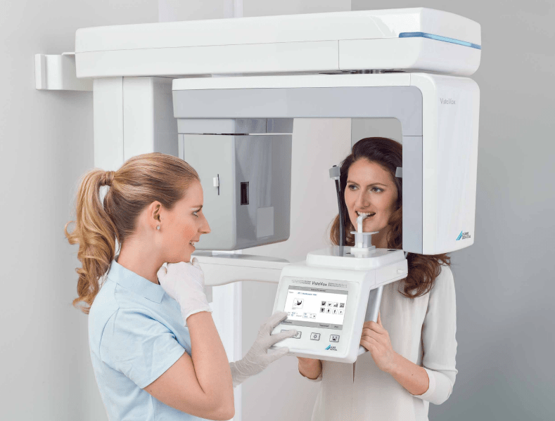
Ideal imaging volume, easy positioning, high image quality: VistaVox S represents a milestone in the field of 3D X-ray systems. Thanks to its unique technology, the 3D images generated with this system cover everything you need for reliable diagnoses, well-founded treatment decisions and convincing patient communication. The S-Pan technology of the VistaVox S generates pinpoint accurate OPG images in best DÜRR DENTAL quality.

The intuitive 8” glass touch display makes operation even more user-friendly. It displays all settings “crystal-clear”. To start with, simply select the required X-ray programme and the correct patient size – this prevents operating errors and ensures that the workflow is optimised.
The special feature of VistaVox S is that its imaging volume is based on the human anatomy, representing precisely the area you need for diagnostics in the dental region.
VistaSoft 3D offers different views for your diagnosis of CBCT images: pano views, transversal layer images (TSA) and multiplanar reconstruction (MPR). Naturally, the VistaVox system includes VistaSoft as an unlimited network licence and a floating licence for three 3D workstations.
With S-Pan technology, the image regions that best correspond to the actual patient anatomy are automatically selected from a large number of parallel layers. The result is an image of impressive clarity, in which you will be able to immediately and effortlessly locate all anatomically relevant structures. Since the reconstruction is aligned to the actual position of the bite, incorrect positioning is compensated for to a certain extent. This saves time for the surgery and prevents the patient from having to have repeat images taken.
In addition to the various CBCT volumes and the 17 panoramic programs, VistaVox S Ceph also offers six modes for all types of cephalometric exposures. The very short scan time starting at just 1.9 seconds helps to avoid motion artefacts and to reduce the radiation dose. The modern high-sensitivity CsI sensors enable excellent image quality.
Metal artefacts are a real challenge for 3D imaging. In the three-dimensional reconstruction, radiopaque objects generate shadows (areas that are displayed completely white) and streaks. Particularly on patients with metal prosthetics, these artefacts make diagnostic work much more difficult. VistaVox MAR eliminates these metal artefacts automatically with the aid of state-of-the-art algorithms and is able to present anatomical structures much better as a result.
With the Comfort Bite, VistaVox S offers an even more stable fixation of the patient, which additionally reduces movement during the exposure.
Thanks to its wider and softer support, it enables more comfortable positioning and is also ideal for edentulous patients
Ideal 3D imaging volume matched to the jaw arch (Ø 130 x 85 mm)
In 2D and 3D thanks to the high-resolution CsI sensor with a pixel size of 49.5 μm
Modern, ergonomic VistaSoftimage processing software
Time-saving cephalometric projections from 1.9 seconds at a low dose


Image detector type
Pixel size (µm)
Scan times (s)
| VistaVox S | VistaVox S Ceph | |
| Image detector type | CsI CMOS Photodiode array | Csl CMOS Photodiode array |
| Pixel size (µm) | 49,5 | 49,5 / 100 |
| Scan times (s) | 2 - 18 | 1,9 - 18 |
| Active sensor surface area (mm) | 135,8 x 36,4 | 135,8 x 36,4 / 157,2 x 16,3 |
| Voltage (kV) | 60 - 99 | 60 - 99 |
| Tube current (mA) | 4 - 16 | 4 - 16 |
| Focal spot (mm) | 0,5 | 0,5 |
| Min. total filtration (mm) | 2,8 | 2,8 |
| 3D volume, child, diagnostic (mm) | Ø 130 x 70 | Ø 130 x 70 |
| 3D volume, adult, diagnostic (mm) | Ø 130 x 85 | Ø 130 x 85 |
| 3D volume, jaw region (mm) | Ø 50 x 50 | Ø 50 x 50 |
| Height (mm) | 1.406 - 2.250 | 1.406 - 2.206 |
| Weight (kg) | 180 | 202 |
| Height adjustment range (mm) | 844 | 800 |
| Width x depth (mm) | 990 x 1.130 | 1.941 x 1.615 |
| Installation (wall-mounted) | ||
| Frequency (Hz) | 50/60 | 50/60 |
| Rated power (VA) | 2.2 | 2.2 |
| Mains voltage (V AC) | 200 - 240 | 200 - 240 |
Image detector type
Pixel size (µm)
Scan times (s)
| VistaVox S | VistaVox S Ceph | |
| Image detector type | CsI CMOS Photodiode array | Csl CMOS Photodiode array |
| Pixel size (µm) | 49,5 | 49,5 / 100 |
| Scan times (s) | 2 - 18 | 1,9 - 18 |
| Active sensor surface area (mm) | 135,8 x 36,4 | 135,8 x 36,4 / 157,2 x 16,3 |
| Voltage (kV) | 60 - 99 | 60 - 99 |
| Tube current (mA) | 4 - 16 | 4 - 16 |
| Focal spot (mm) | 0,5 | 0,5 |
| Min. total filtration (mm) | 2,8 | 2,8 |
| 3D volume, child, diagnostic (mm) | Ø 130 x 70 | Ø 130 x 70 |
| 3D volume, adult, diagnostic (mm) | Ø 130 x 85 | Ø 130 x 85 |
| 3D volume, jaw region (mm) | Ø 50 x 50 | Ø 50 x 50 |
| Height (mm) | 1.406 - 2.250 | 1.406 - 2.206 |
| Weight (kg) | 180 | 202 |
| Height adjustment range (mm) | 844 | 800 |
| Width x depth (mm) | 990 x 1.130 | 1.941 x 1.615 |
| Installation (wall-mounted) | ||
| Frequency (Hz) | 50/60 | 50/60 |
| Rated power (VA) | 2.2 | 2.2 |
| Mains voltage (V AC) | 200 - 240 | 200 - 240 |
With the VistaSoft package you will gain access to powerful AI features that will make your day-to-day work noticeably more effective. In combination with VistaScan IQ image plates, Vistasoft AI enables automatic checking of every X-ray image to detect deterioration in image quality over time – the software will recommend replacement of the image plate where necessary, so that consistently high image quality is ensured.
If you would like further information about Pearl Dental Imaging, please contact us or call the number below