VistaPano S 2.0 and VistaPano S Ceph 2.0

Digital panoramic X-ray device with S-Pan technology

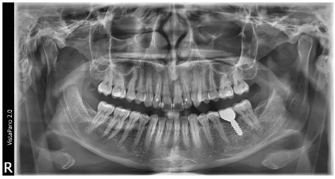
With the VistaPano S 2.0, Dürr Dental has set new standards in the resolution of extraoral images. The 2D panorama X-ray device also impresses with its simple operation and a consistently well thought-out workflow, which is supported by an innovative 8” glass touch display. The excellent imaging performance of the VistaPano S 2.0 is thanks to two innovative technologies. Firstly, the state-of-the-art Csl sensor technology delivers improved picture quality and thus facilitates significantly improved diagnostics. Secondly, S-Pan technology uses the image information from the individual patient image to automatically create a pinpoint-sharp panoramic image of every tooth and jaw position.

Intuitive 8” touchscreen display

All functions at a glance

The intuitive 8” glass touch display makes operation even more user-friendly. It displays all settings “crystal-clear”. To start with, simply select the required X-ray programme and the correct patient size – this prevents operating errors and ensures that the workflow is optimised.

Simple and efficient patient positioning

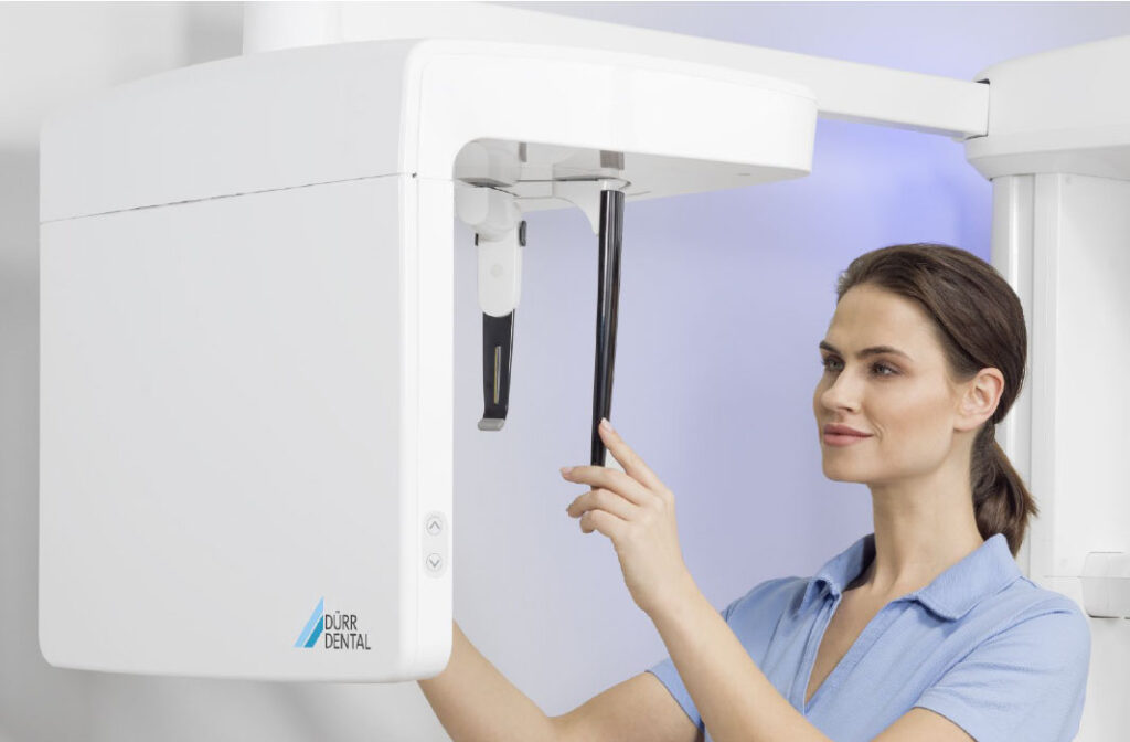
Three laser guide lines mark the exact orientation of Frankfurt horizontal plane, median sagittal plane, and film plane. Positioning without a mirror ensures personal eye contact and facilitates communication with the patient.

Panoramic X-ray programs

With a total of 17 X-ray programs, you are well equipped for every diagnostic requirement. In addition to the standard Panorama program, the VistaPano S 2.0 offers:

  • Half-side recordings of right, left and front
  • 4 child programs: an acquisition mode with a reduced exposure area and a reduction in dose of 45 - 56% without any loss of diagnostic information
  • 5 programs for orthogonal X-ray images
  • 2 programs for temporomandibular imaging
  • 2 programs for sinus X-ray images to display paranasal sinuses

Exceptional image quality

In contrast to the conventional method, S-Pan technology automatically selects the image areas that best represent the patient's individual anatomy from a large number of parallel slices. When the panoramic image is pieced together, deviations from the “average dentition” are taken into account, as is the individual inclination of the teeth. The result is an image of impressive clarity, in which the dentist can effortlessly locate all relevant structures. Since the reconstruction is aligned to the actual position of the bite, incorrect positioning is compensated for to a certain extent. This saves time for the surgery and prevents the patient from having to have repeat images taken.

VistaPano S Ceph 2.0: Improved efficiency – less radiation exposure

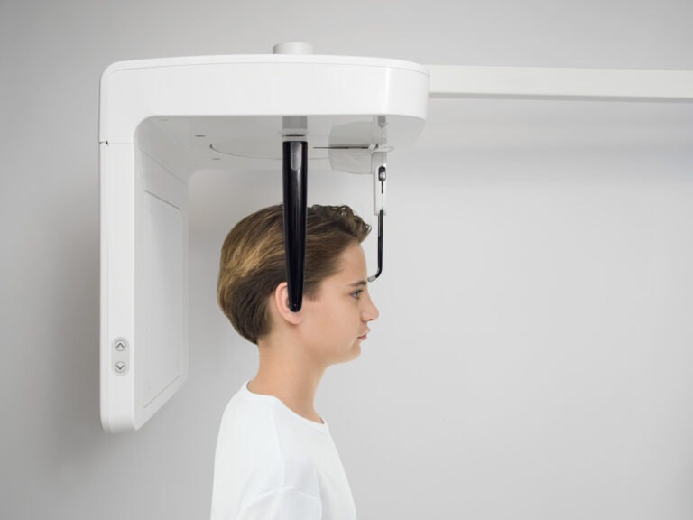
With the new VistaPano S Ceph 2.0, rapid cephalometric images can be taken with outstanding image quality – and with the lowest possible radiation exposure. all thanks to the extremely short scanning time of 1.9 seconds. This reduces the risk of motion fuzziness. A combination of low X-ray dose and short scanning time is especially advantageous with younger orthodontics patients. The two high-end CsI sensors – for panoramic X-ray and Ceph functions – which are already integrated in the unit eliminate the time-consuming switching of the sensor and the risk of damage. All this makes VistaPano S Ceph 2.0 the perfect X-ray solution for orthodontic surgery and for oral & maxillofacial surgery.

Key facts

Compact

Slim design with a small footprint

Fast

Panoramic image acquisition within just 7 seconds / Ceph: Ultra-fast scans within just 2 seconds

Autonomous

8” glass touch display for intuitive operation

Efficient

Outstanding image quality thanks to the latest generation of high-resolution CsI sensors

Cephalometric exposures

Technical Data

CsI:TI Sensor

Image detector type

100

Pixel size (µm)

2,4 - 13,5

Scan times (s)

All Data

CsI:TI Sensor

Image detector type

100

Pixel size (µm)

2,4 - 13,5 (lat.head in fast scan mode: 1,9)

Scan times (s)

All Data
Artificial Intelligence

VistaSoft AI included

With the VistaSoft package you will gain access to powerful AI features that will make your day-to-day work noticeably more effective. In combination with VistaScan IQ image plates, Vistasoft AI enables automatic checking of every X-ray image to detect deterioration in image quality over time – the software will recommend replacement of the image plate where necessary, so that consistently high image quality is ensured.

VistaScan Combi Documentation

Free Consultation

Get In Touch Today

If you would like further information about Pearl Dental Imaging, please contact us or call the number below

Phone: 0116 275 9995
Address: Pearl Dental Imaging, 69 Enderby Road, Blaby, Leicester, Leicestershire, LE8 4GD